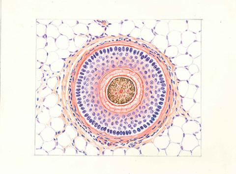
稀突起神経膠細胞 (犬,大脳皮質) リオホルテガ鍍銀法 ×200
Cite
"稀突起神経膠細胞 (犬,大脳皮質) リオホルテガ鍍銀法 ×200" owned by Nagoya University Museum, retrieved from Human Tissue Illustrations(https://da.adm.thers.ac.jp/en/item/n011-20230901-00086)
Copy
ID
NUM-Lp002-086
Title
稀突起神経膠細胞 (犬,大脳皮質) リオホルテガ鍍銀法 ×200
Source
Nagoya University Graduate School of Medicine
Note
名古屋大学医学部 解剖学教室 旧蔵 (箱1)
Material type
StillImage (PDF)
Original Owner
Nagoya University Museum
Image
colour
Collection
Human Tissue Illustrations
This is an image of " Human Tissue Illustrations of Nagoya University", a booklet containing 160 of the 435 tissue drawings by Shiro Kido (1938-1969), a technician in the Department of Functional Histology (Second Department of Anatomy) and a painter.
related items
大神経膠細胞 (犬,大脳皮質) リオホルテガ鍍銀法 ×200
Human Tissue Illustrations Nagoya University Museum
小神経膠細胞 (犬,大脳垂直断) リオホルテガ鍍銀法 ×200
Human Tissue Illustrations Nagoya University Museum
多極神経細胞 (犬, 大脳皮質) ヴィルショウスキー染色 ×450
Human Tissue Illustrations Nagoya University Museum
自由神経終末 (豚,鼻先上皮) 鈴木鍍銀法 ×200
Human Tissue Illustrations Nagoya University Museum
神経原線維 犬 脊髄 神経細胞 ビールショウスキー染色 ×200
Human Tissue Illustrations Nagoya University Museum
ファーター・パチニ小体 (人,手指皮下細胞内) 鈴木鍍銀法 ×200
Human Tissue Illustrations Nagoya University Museum
