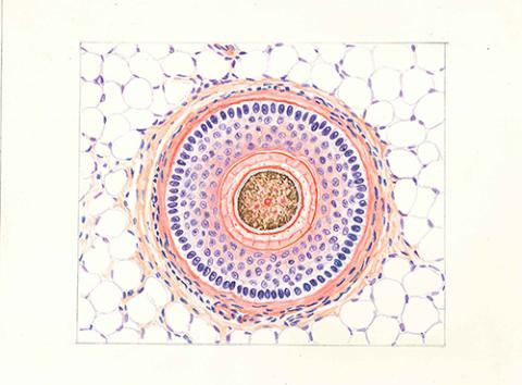
This image archive is part of the collection of human tissue drawings transferred from the Nagoya University Graduate School of Medicine to the Nagoya University Museum in 2010, and was published in the booklet "Nagoya University Human Tissue Drawings" (Author: Shiro Kido, Editing authors: Hiroshi Kiyama and Seiji Kadowaki) produced as part of the 150th anniversary commemoration project of Nagoya University Medical School. (Author: Shiro Kido, Editing authors: Hiroshi Kiyama and Seiji Kadowaki). There are 435 original histological drawings in total, and they are stored at the Nagoya University Museum as a series of material number NUM-Lp002.
Background of the booklet "Nagoya University Human Tissue Drawings"
Prior to my appointment at Nagoya University, I had been involved in teaching histology and gross anatomy at three different universities. When I was appointed to the Department of Functional Histology (Anatomy II) at Nagoya University in 2011, I was greatly impressed by the excellence of the tissue specimens for student practice. Moreover, I was surprised that slides were used for practical training that allowed students to find almost the same locations as the photographs and illustrations in "Histology" (Nanzando), which is used as a textbook. Furthermore, there were many old histological diagrams that were partially used in textbooks, etc. These are undoubtedly wonderful assets of Nagoya University, and at the same time, I thought how lucky the students of Nagoya University are.
I learned that these diagrams were drawn by the painter Mr. Shiro Kido (Nagoya University, 1938-1969) under the guidance of Professor Kintaro Tokari (Professor of Anatomy II, 1933-1960), who was a professor four generations before me, and that the textbook "Histology" was published by Nanzando in 1954, using some of these diagrams. I learned that the textbook "Histology" was published by Nanzando in 1954, using some of these diagrams.
In 1972, after his retirement, Mr. Shiro Kido received the 13th CBC Club Cultural Award, and in his commendation, he stated, "In medical education, there have been few cases in which the human eye has developed areas where the microscope could not be used to create illustrations. But you are the one who has done it, who has amazed the society, and who has earned its gratitude. These are the words of Mr. Shiro Kido.
Professor Yasuo Sugiura and Associate Professor Shinya Kobayashi of the Department of Functional Histology (Anatomy II) were responsible for the transfer and preservation of the tissue charts left by Dr. Shiro Kido to the Nagoya University Museum as a unique asset for medical education. Both professors collaborated with Dr. Masumi Nozaki, then a researcher at the Nagoya University Museum, to digitize the histological maps, and the originals were transferred to the Nagoya University Museum in 2010. After that, we were looking for funding to publish them, but we were unable to make any progress. In 2021, at a banquet, I spoke with Dr. Kenji Kadomatsu, then Dean of the Graduate School, and Dr. Hiroshi Kimura, current Dean of the Graduate School, who was the chairman of the 150th anniversary commemorative project of the founding of the Nagoya University School of Medicine, and they suggested that we make this one of the commemorative projects for the 150th anniversary of the founding. He gave us permission to use part of his donation for this publication. This led to a discussion of publishing the illustrated book at once, and we decided to edit it together with Dr. Seiji Kadowaki of the Nagoya University Museum, who was planning an exhibition on Shiro Kido for another 150th anniversary of the foundation. Of the 435 original drawings that are still in existence, 160 have been compiled into this "Nagoya University Human Histology Scroll". This illustrated catalog has both academic and artistic value, and on the occasion of the 150th anniversary of the founding of the Nagoya University School of Medicine, it is an asset of Nagoya University that should be passed on for many years to come.
Finally, I would like to express my deepest gratitude to Dr. Yasuo Sugiura, Dr. Shinya Kobayashi, and Dr. Masumi Nozaki, whose enthusiasm laid the foundation for this publication, and to Fumiko Asano, a technical staff member currently in charge of histology, and Ayami Watanabe, a researcher at the Nagoya University Museum, for their efforts in the editing process. I would also like to acknowledge the efforts of Fumiko Asano, currently a technical staff member in charge of histology, and Ayami Watanabe, a researcher at the Nagoya University Museum, in the editing process.
April 2022
Department of Functional Histology (Anatomy II), Nagoya University Graduate School of Medicine
Hiroshi Kiyama
Artist Shiro Kido drew human tissue illustrations. 1900 – 1986
Shiro Kido is a Western-style painter born in Atsumi-gun, Aichi Prefecture. After studying at the Kawabata School of Painting in Koishikawa, Tokyo, he established a studio in Nagoya. He painted many nature and landscape scenes, including camellias on the beach at his hometown of Cape Irago, and held solo exhibitions at the Maruzen Gallery in Nagoya. Some of Kido's works are in the collection of the Atsumi Local History Museum (Tahara City Museum, 2012).
In addition to his activities as a painter, Kido began working as a technical officer in the Department of Anatomy at Aichi Medical University (now Nagoya University School of Medicine) in 1938. At the request of Professor Kintaro Tokari of the School of Medicine, he drew tissue charts.
Using his talent as a painter, Mr. Kido drew the colorful tissue charts with his brush, which could not be expressed by photographs. It was a precise tissue chart unparalleled anywhere in the world. According to Kido's daughter, who watched him work as a child, she "couldn't help but wonder" as he looked through a microscope with one eye while his right hand moved animatedly to draw the diagram (CBC Club, 1985). According to Kido, it took four to five days to draw a single figure for the longest one, and about one day for the shortest one (CBC Club, ed. 1972).
Mr. Kido drew approximately 500 organizational charts over a period of 30 years or so, until he retired from Nagoya University in 1969. During that time, he was drafted into the military from the Sino-Japanese War to the Pacific War, and experienced harsh conditions as a conscript.
Kido's watercolors, which accurately depict microscopic images of tissue specimens, were published in the textbook "Histology" (1954, Nanzando) by Professor Kintaro Tokari. In the preface to this book, Professor Tokari wrote the following
It is no exaggeration to say that without understanding histology, it is impossible to understand basic anatomy and pathology, let alone the true essence of clinical medicine. The tissue specimens used for the illustrations were faithfully depicted by painter Kido from specimens that had been used for the display and practice of students in the Department of Anatomy, Nagoya University School of Medicine for the past 30 years. The rest are based on human bodies.
The tissue drawings by Shiro Kido contributed to the education of many medical students throughout Japan when they were published as illustrations in Professor Tokari's "Histology.
In 1972, Shiro Kido received the 13th CBC Club Cultural Award (Kuchinashi Chapter) for his contribution to medical education by raising anatomical tissue charts to a world-class level, as well as for his efforts to improve local culture.
The 435 original drawings of this organizational chart are now housed in the Nagoya University Museum, where they are used as exhibits and other materials to convey the history of medical education at Nagoya University.
Finally, Mr. Kikuji Sakai, Secretary General of the CBC Club, provided valuable references on Shiro Kido in preparing this chart, and we have reproduced the photographs of Professor Kintaro Tokari and Shiro Kido in this book. We would like to express our gratitude to him.
Nagoya University Museum
Seiji Kadowaki
References
CBC Club (ed.) (published February 25, 1972), CBC Club News, No. 137,
CBC Club (ed.) (published February 1, 1985), Mokimoku ichigugei ichinou: it's you, CBC Club Cultural Award no 25 nen, Chubu-Nippon Broadcasting Co.
Tahara City Museum (ed., published October 27, 2012), "Tahara City Atsumi Local History Museum, 2012 Autumn Special Exhibition: Painters Related to the Local Area," Tahara City Museum
Tokari, Kintaro (published in 1954), "Historiography", Nanzando
Please visit the Nagoya University Museum website for more information on use.
The thumbnail image is from "毛 (人,頭皮横断) 上部 H-E染色 ×200" owned by Nagoya University Museum, retrieved from Human Tissue Illustrations(https://da.adm.thers.ac.jp/en/item/n011-20230901-00201)
